


Special Offers
100% Performance Guaranteed
Key Specifications Table
| Species Reactivity | Key Applications | Host | Format | Antibody Type |
|---|---|---|---|---|
| Vrt | ICC, IP, WB | Ch | Purified | Polyclonal Antibody |
| Description | |
|---|---|
| Catalogue Number | AB16901 |
| Brand Family | Chemicon® |
| Trade Name |
|
| Description | Anti-Green Fluorescent Protein Antibody |
| Alternate Names |
|
| Background Information | The Green Fluorescent Protein (GFP) from the jellyfish Aequorea victoria is used as a fluorescent indicator for monitoring gene expression in a variety of cellular systems, including living organisms and fixed tissues. Unlike other bioluminescent reporters, GFP fluoresces in the absence of substrates, cofactors, or other intrinsic or extrinsic proteins. Purified GFP is a 27 kDa monomer consisting of 238 amino acids and emits green light (emission maximum at 509 nm) when excited with blue or UV light. |
| Product Information | |
|---|---|
| Format | Purified |
| Control |
|
| Presentation | Purified chicken immunoglobulin in TBS, pH 7.5 with 0.02% sodium azide. |
| Quality Level | MQ100 |
| Applications | |
|---|---|
| Application | This Anti-Green Fluorescent Protein Antibody is validated for use in IC, IP, WB for the detection of Green Fluorescent Protein. |
| Key Applications |
|
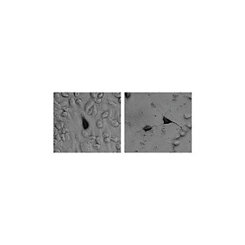
| Application Notes | Immunoblotting: AB16901 detects a band of the proper molecular weight (30kDa) in lysates from E.coli expressing GFP but not in E.coli that do not express or in lysates prepared from E.coli containing the GFP-expressing plasmid but not induced, detect GFP in lysates from COS or primary neurons or fibroblast cells transfected with plasmids directing expression GFP or a tau-GFP fusion protein. Did not detect any signal when 50μg of protein from a rat brain lysate (negative control) was fractionated on the SDS-PAGE. Titers were 10-20,000 when detected with an anti-chicken horseradish peroxidase-conjugated secondary by ECL or by a colorimetric assay using DAB. Immunocytochemistry: GFP can be detected in cells that are transfected with a plasmid directing the expression of either GFP or a GFP-Fusion protein. Cells that do not express GFP exhibit no detectable staining. AB binding was determined using HRP conjugated-anti chicken secondary Abs and visualized using DAB. The anti-GFP Abs were used at 1-2,500-5,000 fold dilution. Cells were fixed using 4% formaldehyde in PBS (pH7.4). For immunocytochemistry, the cultures were fixed in 4% formaldehyde in PBS pH7.4, for 20 minutes at room temperature. The cells are permeabilized with 0.2% Triton X-100 and 2% serum (corresponding to the aminal in which the secondary antibody was raised) in PBS for 20 minutes at room temperature. Cell cultures are then incubated with primary antisera in PBS containing 2% serum (as above), at 4°C, overnight. Antibody binding is detected using an avidin/biotin/peroxidase detection system. Cultures were washed four times (10 minutes each) between steps, and reaction product is developed with a 0.05% diaminobenzidine / 0.02% hydrogen peroxide solution on ice for 8-10 minutes. AB16901 has been used to detect GFP in primary neurons and zebrafish fibroblasts. Immunoprecipitation: 5μg antibody per no more than 500μl of cell lysate. It is important to use a rabbit anti-chicken secondary for capture or anti-chicken beads as Chicken IgG will not react with protein A or protein G. Optimal working dilutions must be determined by the end user. |
| Biological Information | |
|---|---|
| Immunogen | A recombinant green fluorescent protein (GFP) containing a 6-his tag, was highly purified by affinity chromatography on Nickel column followed by chromatography on a P-100 gel filtration column. The resulting product was present as only a singl |
| Concentration | Please refer to the Certificate of Analysis for the lot-specific concentration. |
| Host | Chicken |
| Specificity | AB16901 is made against highly-purified native GFP from Aequorea victoria. It is reactive with GFP from both native and recombinant sources. |
| Species Reactivity |
|
| Antibody Type | Polyclonal Antibody |
| Purification Method | Ammonium Sulfate Precipitation |
| UniProt Number |
|
| Molecular Weight | MW for GFP is 27 kDa. Dependent upon the molecular weight of the fusion protein being detected. |
| Product Usage Statements | |
|---|---|
| Usage Statement |
|
| Storage and Shipping Information | |
|---|---|
| Storage Conditions | The undiluted antibody solution is stable for approximately 6 months stored between 2 and 8°C. During shipment, small volumes of product will occasionally become entrapped in the seal of the product vial. For products with volumes of 200μL or less, we recommend gently tapping the vial on a hard surface or briefly centrifuging the vial in a tabletop centrifuge to dislodge any liquid in the containers cap. |
| Packaging Information | |
|---|---|
| Material Size | 250 µg |