


Special Offers
100% Performance Guaranteed
Key Specifications Table
| Species Reactivity | Key Applications | Host | Format | Antibody Type |
|---|---|---|---|---|
| R, A | IH(P), ELISA | Gp | Serum | Polyclonal Antibody |
| Description | |
|---|---|
| Catalogue Number | AB175 |
| Brand Family | Chemicon® |
| Trade Name |
|
| Description | Anti-GABA Antibody |
| Background Information | GABA (gamma-aminobutyric acid) is the major inhibitory neurotransmitter in the central nervous system that inhibits the generation of the action potential of the neuron. It is involved in the pathogenesis of certain neurological and psychiatric disorders. GABA is produced from glutamic acid in a reaction catalyzed by glutamic acid decarboxylase. In this reaction pyridoxal phosphate serves as a co-factor. GABA interacts with the GABAA and GABAB receptors, which are widely distributed throughout the nervous system and in a variety of cell types. They differ in their pharmacological, electrophysiological, and biochemical properties. GABAA-receptor complex mediates an increase in membrane conductance with an equilibrium potential near the resting level of −70 mV. This conductance increase often is accompanied by a membrane hyperpolarization, which later results in a reduction in the probability of action potential initiation. The reduction in membrane resistance is accomplished by the GABA-dependent facilitation of Cl− ion influx. GABAB receptors are coupled indirectly to K+ channels. When activated, GABAB receptors reduce Calcium ion conductance and inhibit cAMP production via intracellular mechanisms mediated by G-proteins. GABAB receptors are known to mediate both postsynaptic and presynaptic inhibition. |
| Product Information | |
|---|---|
| Format | Serum |
| Control |
|
| Presentation | Guinea Pig polyclonal BSA-depleted antiserum with 0.05% sodium azide. |
| Quality Level | MQ100 |
| Applications | |
|---|---|
| Application | This Anti-GABA Antibody is validated for use in Immunohistochemistry (Paraffin) and Enzyme Immunoassay (ELISA) for the detection of GABA. |
| Key Applications |
|
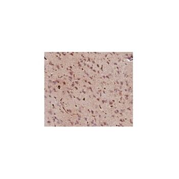
| Application Notes | Immunohistochemistry Analysis: A 1:500 dilution from a representative lot detected GABA in rat brain tissue. ELISA Analysis: A representative lot from an independent laboratory detected GABA in a competitive ELISA. Immunohistochemistry Protocol: Tissues fixed with 4% paraformaldehyde and 0-0.5% glutaraldehyde gives good results. Glutaraldehyde is required for antibody reactivity. 1) Tissue is fixed with 4% paraformaldehyde, 0-0.5% glutaraldehyde, 0.5% potassium dichromate in 0.1M phosphate buffer at pH 6.5. 2) Tissue is post-fixed overnight, vibratome sectioned in 50 mm and incubated in 0.05M Tris buffer, pH 6.5 for three hours. 3) Sections are incubated for 18-24 hours in AB175 diluted in PBS containing 0.1% sodium azide, 0.2% Triton X-100 and 1% normal goat serum. 4) Fluorescein conjugated antibody or ABC system may be used as the secondary reagent. Note: Without colchicine pretreatment well-stained cell bodies are visible in the cerebral cortex, cerebrallar cortex, superior colliculus and some brainstem raphe. With colchicine pretreatment, additional cell body staining is present in the interpeduncular nucleus and the dorsal column nuclei. |
| Biological Information | |
|---|---|
| Immunogen | BSA-conjugated GABA-Glutaraldehyde |
| Concentration | Variable |
| Host | Guinea Pig |
| Specificity | Recognizes GABA. Staining was blocked by preabsorbing with 100 μM GABA conjugated to glutaraldehyde. 500 μM of similar conjugations of glutamic acid, glutamate and taurine failed to block staining. |
| Species Reactivity |
|
| Species Reactivity Note | Demonstrated to react with Rat. GABA is a neurotransmitter synthesized in all species, and is therefore expected to react to all species. |
| Antibody Type | Polyclonal Antibody |
| Purification Method | Depleted serum |
| Product Usage Statements | |
|---|---|
| Quality Assurance | Evaluated by Immunohistochemistry in rat brain tissue. Immunohistochemistry Analysis: A 1:500 dilution of this antibody detected GABA in rat brain tissue. |
| Usage Statement |
|
| Storage and Shipping Information | |
|---|---|
| Storage Conditions | Stable for 1 year at -20°C from date of receipt. Handling Recommendations: Upon receipt and prior to removing the cap, centrifuge the vial and gently mix the solution. Aliquot into microcentrifuge tubes and store at -20°C. Avoid repeated freeze/thaw cycles, which may damage IgG and affect product performance. |
| Packaging Information | |
|---|---|
| Material Size | 50 µL |