


Special Offers
100% Performance Guaranteed
Key Specifications Table
| Species Reactivity | Key Applications | Host | Format | Antibody Type |
|---|---|---|---|---|
| H, M, R, B, Po | ICC, IHC, IH(P), WB | Ch | Affinity Purified | Polyclonal Antibody |
| Description | |
|---|---|
| Catalogue Number | AB5541 |
| Replaces | 04-1031; 04-1062 |
| Brand Family | Chemicon® |
| Trade Name |
|
| Description | Anti-Glial Fibrillary Acidic Protein Antibody |
| Alternate Names |
|
| Background Information | Glial fibrillary acidic protein (GFAP) is a class-III intermediate filament. Type III intermediate filaments contain three domains, the most conserved of which is the rod domain. The specific sequence for this region of the protein may differ between the different intermediate filament genes for type III proteins, but the structure of the protein is highly conserved. GFAP is the main constituent of intermediate filaments in astrocytes and serves as a cell specific marker that distinguishes differentiated astrocytes from other glial cells during the development of the central nervous system. GFAP is also found in the lens epithelium, Kupffer cells of the liver, in some cells in salivary tumors and has been reported in erythrocytes. |
| Product Information | |
|---|---|
| Format | Affinity Purified |
| Control |
|
| Presentation | Purified chicken polyclonal in buffer containing PBS containing 0.02% sodium azide. |
| Quality Level | MQ100 |
| Applications | |
|---|---|
| Application | This Anti-Glial Fibrillary Acidic Protein Antibody is validated for use in IC, IH, IH(P), WB for the detection of GFAP. |
| Key Applications |
|
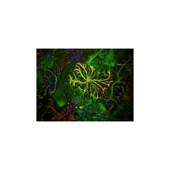
| Application Notes | Immunohistochemistry(paraffin): Representative images from a previous lot. Pictures of Optimal Staining With Citrate Buffer Epitope Retrieval: Human Brain. Immunocytochemistry: 1:200-1:1,000 on cells in tissue culture fixed for one minute in 3.7% formalin and one minute in -20°C methanol. Western blot: 1:100-1:500 using ECL. Recognizes a band of 55 kDa. May also react with a band of 50 kDa. Immunohistochemistry: 1:100-1:500 on frozen tissue sections. Suggested fixative is 4% paraformaldehyde. Optimal working dilutions must be determined by the end user. STAINING PATTERN: The antibody stains sharply defined cytoplasmic filaments of astroglia cells in tissue culture. Stains processes of astrocytes in sections of brain tissues. |
| Biological Information | |
|---|---|
| Immunogen | Purified bovine GFAP. |
| Concentration | Please refer to the Certificate of Analysis for the lot-specific concentration. |
| Host | Chicken |
| Specificity | Glial fibrillary acidic protein (GFAP). Reacts with both native and recombinant protein. |
| Species Reactivity |
|
| Species Reactivity Note | Human, rat, and mouse. Expected to cross-react with bovine and porcine. Other species have not yet been tested. |
| Antibody Type | Polyclonal Antibody |
| Entrez Gene Number |
|
| Entrez Gene Summary | This gene encodes one of the major intermediate filament proteins of mature astrocytes. It is used as a marker to distinguish astrocytes from other glial cells during development. Mutations in this gene cause Alexander disease, a rare disorder of astrocytes in the central nervous system. An additional transcript variant has been described, but its full length sequence has not been determined. |
| Gene Symbol |
|
| Purification Method | ImmunoAffinity Purified |
| UniProt Number |
|
| UniProt Summary | FUNCTION: SwissProt: P14136 # GFAP, a class-III intermediate filament, is a cell- specific marker that, during the development of the central nervous system, distinguishes astrocytes from other glial cells. SIZE: 432 amino acids; 49880 Da SUBUNIT: Isoform 3 interacts with N-terminus of PSEN1. SUBCELLULAR LOCATION: Cytoplasm. Note=Associated with intermediate filaments. DISEASE: SwissProt: P14136 # Defects in GFAP are a cause of Alexander disease [MIM:203450]. Alexander disease is a rare disorder of the central nervous system. It is a progressive leukoencephalopathy whose hallmark is the widespread accumulation of Rosenthal fibers which are cytoplasmic inclusions in astrocytes. The most common form affects infants and young children, and is characterized by progressive failure of central myelination, usually leading to death usually within the first decade. Infants with Alexander disease develop a leukoencephalopathy with macrocephaly, seizures, and psychomotor retardation. Patients with juvenile or adult forms typically experience ataxia, bulbar signs and spasticity, and a more slowly progressive course. SIMILARITY: SwissProt: P14136 ## Belongs to the intermediate filament family. ... |
| Molecular Weight | 55 kDa |
| Product Usage Statements | |
|---|---|
| Quality Assurance | Routinely evaluated by Western Blot on Mouse Brain lysates. Western Blot Analysis: 1:1000 dilution of this lot detected glial fibrillary acidic protein on 10 μg of Mouse Brain lysates. |
| Usage Statement |
|
| Storage and Shipping Information | |
|---|---|
| Storage Conditions | Stable for 1 year at -20°C in undiluted aliquots from date of receipt. Do not store in a self-defrosting freezer. Handling Recommendations: Upon first thaw, and prior to removing the cap, centrifuge the vial and gently mix the solution. Aliquot into microcentrifuge tubes and store at -20°C. Avoid repeated freeze/thaw cycles, which may damage IgG and affect product performance. |
| Packaging Information | |
|---|---|
| Material Size | 50 µL |