


Special Offers
100% Performance Guaranteed
Key Specifications Table
| Species Reactivity | Key Applications | Host | Format | Antibody Type |
|---|---|---|---|---|
| H, M, R | IH(P), WB | Rb | Affinity Purified | Polyclonal Antibody |
| Description | |
|---|---|
| Catalogue Number | AB9260 |
| Brand Family | Chemicon® |
| Trade Name |
|
| Description | Anti-Ki-67 Antibody |
| Background Information | Proliferation marker protein Ki-67 (UniProt: P46013; also known as Antigen identified by monoclonal antibody Ki-67, KI-67, Ki67) is encoded by the MKI67 gene (Gene ID: 4288) in human. Ki67 antigen is the prototypic cell cycle related nuclear protein, expressed by proliferating cells in all phases of the active cell cycle (G1, S, G2 and M phase). In the G1 phase it is predominantly localized in the perinucleolar region and in the later phases it is also detected throughout the nuclear interior, being predominantly localized in the nuclear matrix. Its levels are shown to be the highest in the G2 phase and during mitosis. Ki67 is routinely used as a marker of cell cycling and proliferation. It is required to maintain individual mitotic chromosomes dispersed in the cytoplasm following nuclear envelope disassembly. It is shown to associates with the surface of the mitotic chromosome, the perichromosomal layer, and covers a substantial fraction of the chromosome surface and prevents chromosomes from collapsing into a single chromatin mass by forming a steric and electrostatic charge barrier. It binds DNA, with a preference for supercoiled DNA and AT-rich DNA. Ki67 undergoes phosphorylation and is shown to be hyperphosphorylated in mitosis phase. The hyperphosphorylated form fails to bind DNA. Two isoforms of Ki67 have been described that are produced by alternative splicing. |
| Product Information | |
|---|---|
| Format | Affinity Purified |
| Control |
|
| Presentation | Purified rabbit polyclonal in buffer containing 10 mm PBS, pH 7.6 containing 1% BSA and 0.099% sodium azide. |
| Quality Level | MQ100 |
| Applications | |
|---|---|
| Application | Detect Ki-67 with Anti-Ki-67 Antibody (Rabbit Polyclonal Antibody), that has been shown to work in IHC(P) & WB. |
| Key Applications |
|
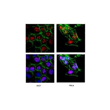
| Application Notes | Immunocytochemistry: Confolcal fluorescent analysis of A431 cells using AB9260 rabbit pAB. Actin filaments were labeled with Alexa Fluor 488 -Phalloidin. Nuclear is stained with DAPI. (Nuclear staining). Immunohistochemistry: A representative lot of this antibody immunostained paraffin-embedded rat testis tissue sections. (Karaca, T., et Al. (2018). Iran. J. Vet. Res. 10(4); 255-261). Immunohistochemistry: A representative lot of this antibody immunostained frozen mouse brain tissue sections. (Zhang, H., et Al. (2019). eLife. 8; e45303). Western Blot Analysis: A previous lot of this antibody was used in western blot. Optimal working dilutions must be determined by the end user. |
| Biological Information | |
|---|---|
| Immunogen | Synthetic peptide from human Ki-67 |
| Concentration | Please refer to the Certificate of Analysis for the lot-specific concentration. |
| Host | Rabbit |
| Specificity | Recognizes Ki-67 |
| Species Reactivity |
|
| Species Reactivity Note | Human, mouse and rat. |
| Antibody Type | Polyclonal Antibody |
| Entrez Gene Number |
|
| Gene Symbol |
|
| Purification Method | ImmunoAffinity Purified |
| UniProt Number |
|
| UniProt Summary | FUNCTION: SwissProt: P46013 # Thought to be required for maintaining cell proliferation. SIZE: 3256 amino acids; 358694 Da SUBUNIT: Binds through the FHA domain to MKI67IP. SUBCELLULAR LOCATION: Nucleus. Note=Predominantly localized in the G1 phase in the perinucleolar region, in the later phases it is also detected throughout the nuclear interior, being predominantly localized in the nuclear matrix. In mitosis, it is present on all chromosomes. DEVELOPMENTAL STAGE: Expression of this antigen occurs preferentially during late G1, S, G2 and M phases of the cell cycle, while in cells in G0 phase the antigen cannot be detected. SIMILARITY: SwissProt: P46013 ## Contains 1 FHA domain. |
| Molecular Weight | Predicted: 359 kDa. MW may be observed at ~345 or 395 kDa |
| Product Usage Statements | |
|---|---|
| Quality Assurance | Immunohistochemistry(paraffin): Representative lot data. This antibody was used on formalin fixed, paraffin embedded tissue at 1:300 dilution for 10 minutes at room temperature. Staining of formalin-fixed tissues requires boiling of tissues in 10 mm citrate buffer, pH 6.0 for 10 minutes followed by cooling at room temperature for 20 minutes. |
| Usage Statement |
|
| Storage and Shipping Information | |
|---|---|
| Storage Conditions | Stable for 1 year at 2-8ºC from date of receipt. |
| Packaging Information | |
|---|---|
| Material Size | 500 µL |