


Special Offers
100% Performance Guaranteed
Key Specifications Table
| Species Reactivity | Key Applications | Host | Format | Antibody Type |
|---|---|---|---|---|
| M, R | WB, IF | Gp | Serum | Polyclonal Antibody |
| Description | |
|---|---|
| Catalogue Number | ABN83 |
| Description | Anti-Vesicular Nucleotide Transporter (VNUT Antibody) |
| Alternate Names |
|
| Background Information | Solute carrier family 17 member 9 (UniProt; also known as Vesicular nucleotide transporter, VNUT) is encoded by the Slc17a9 (also known as Vnut) gene (Gene ID 228993) in murine species. VNUT is an anion transporter family member that mediates the active accumulation of nucleotides (i.e., ATP, ADP, and UTP) into secretory vesicles, as well as the exocytosis of ATP from various tissue types. VNUT is expressed by tyrosine hydroxylase-/TH-positive dopaminergic (DA) amacrine/interplexiform cells (IPCs) in the retina, as well as by both DA and non-DA neurons in the midbrain. In addition to the 447-amino acid Slc17a9 transcript (NM_183161), putative protein coding isoforms based on automated computational analysis have been predicted (X1, XM_006500589; X2, XM_006500590; X3, XM_006500591). Two protein bands of sizes ~75 and ~50 kDa are identified in mouse retinal and cortical tissues by immunoblotting, with the 75 kDa band being the 447-a.a. canonical VNUT isoform and the ~50 kDa band likely representing isoform X1/X2. In addition, a ~60 kDa form is also reported, representing either another spliced form or less glycosylated form. Murine VNUT is a 11-transmembrane (a.a. 40-60, 74-94, 103-123, 129-149, 169-189, 192-212, 252-272, 287-307, 327-347, 380-400, 413-433) vesicular protein, having an extacellular/vacuolar N-terminal end (a.a. 1-39) and a cytoplasmic C-terminal tail (a.a. 434-447) with 5 cytoplasmic and 5 extacellular/vacuolar loops in between. |
| Product Information | |
|---|---|
| Format | Serum |
| Presentation | Guinea pig polyclonal serum with 0.05% sodium azide. |
| Quality Level | MQ100 |
| Applications | |
|---|---|
| Application | This Anti-Vesicular Nucleotide Transporter (VNUT antibody is validated for use in WB, IF for the detection of Vesicular Nucleotide Transporter (VNUT. |
| Key Applications |
|
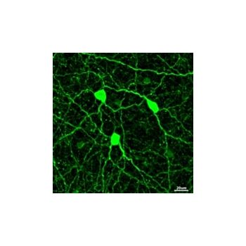
| Application Notes | Immunofluorescence Analysis: A 1:1000 dilution from a representative lot immunostained amacrine/interplexiform cell (IPC) type by fluorescent immunohistochemistry staining of 4% paraformaldehyde-fixed, OCT-embedded mouse and rat vertical retinal cryosections, as well as of flatmounted mouse and rat retinae (Courtesy of Dr. Erica Fletcher, The University of Melbourne, Australia). Western Blotting Analysis: A 1:500 dilution from a representative lot detected purified full-length recombinant VNUT, as well as endogenous VNUT in mouse retina extract (Courtesy of Dr Erica Fletcher, The University of Melbourne, Australia). Immunofluorescence Analysis: A 1:1000 dilution from a representative lot immunostained VNUT-positive neurons in the substantia nigra (SN) by fluorescent immunohistochemistry staining of 4% paraformaldehyde-fixed, free-floating mouse brain cryosections (Courtesy of Dr. Erica Fletcher, The University of Melbourne, Australia). Immunofluorescence Analysis: A representative lot immunostained tyrosine hydroxylase-/TH-positive amacrine/interplexiform cell (IPC) type in the inner nuclear layer (INL) by fluorescent immunohistochemistry staining of 4% paraformaldehyde-fixed, OCT-embedded mouse and rat vertical retinal cryosections, as well as flatmounted mouse retina (Ho, T., et al. (2015). Front. Cell. Neurosci. In Press). Immunofluorescence Analysis: A representative lot immunostained tyrosine hydroxylase-/TH-positive dopaminergic neurons in the substantia nigra (SN) and ventral tegmental area (VTA) by fluorescent immunohistochemistry staining of 4% paraformaldehyde-fixed, free-floating mouse brain cryosections (Ho, T., et al. (2015). Front. Cell. Neurosci. In Press). Western Blotting Analysis: A representative lot detected the ~75 kDa and ~50 kDa X1/X2 VNUT isoforms in murine retinal and cortical homogenates, while no VUNT expression was detected in kidney homogenate. Immunogen peptide blocking abolished target bands detection (Ho, T., et al. (2015). Front. Cell. Neurosci. In Press). |
| Biological Information | |
|---|---|
| Immunogen | KLH-conjugated linear peptide corresponding to a sequence from the second extracellular loop of mouse Vesicular Nucleotide Transporter (VNUT). |
| Epitope | Second extracellular loop. |
| Concentration | Please refer to lot specific datasheet. |
| Host | Guinea Pig |
| Specificity | This guinea pig polyclonal antiserum detected the ~75 kDa, ~60 kDa, and the ~50 kDa (X1/X2 VNUT isoforms) VNUT target bands in murine retinal and cortical homogenates. Immunogen peptide blocking abolished target bands detection. |
| Species Reactivity |
|
| Antibody Type | Polyclonal Antibody |
| Entrez Gene Number |
|
| Gene Symbol |
|
| Purification Method | Unpurified |
| UniProt Number |
|
| Molecular Weight | ~75/60/50 kDa observed. Target band size appears larger than the calculated molecular weight of 48.48 kDa due to glycosylation. Uncharacterized band(s) may appear in some lysates. |
| Product Usage Statements | |
|---|---|
| Quality Assurance | Evaluated by Western Blotting in mouse brain tissue lysate. Western Blotting Analysis: A 1:500 dilution of this antibody detected Vesicular Nucleotide Transporter (VNUT) in 10 µg of mouse brain tissue lysate. |
| Usage Statement |
|
| Storage and Shipping Information | |
|---|---|
| Storage Conditions | Stable for 1 year at -20°C from date of receipt. Handling Recommendations: Upon receipt and prior to removing the cap, centrifuge the vial and gently mix the solution. Aliquot into microcentrifuge tubes and store at -20°C. Avoid repeated freeze/thaw cycles, which may damage IgG and affect product performance. |
| Packaging Information | |
|---|---|
| Material Size | 100 µL |