We use cookies to make your experience better. To comply with the new e-Privacy directive, we need to ask for your consent to set the cookies. Learn more.

Cell Signaling Nut (C52b1) Rabbit mAb
List Price
$342.39
Your Price
$342.39
Cell Signaling Nut (C52b1) Rabbit mAb - CSIG (Additional S&H or Hazmat Fees May Apply)
NETA PART:
CSIG-3625S
MFG.PART:
3625S
UNSPSC:
12352203
Manufacturer:
Cell Signaling


| Size | 100 µl |
| Reactivity | H R |
| Sensitivity | Endogenous |
| Molecular Weight (kDa) | 150 |
| Source/Isotype | Rabbit IgG |
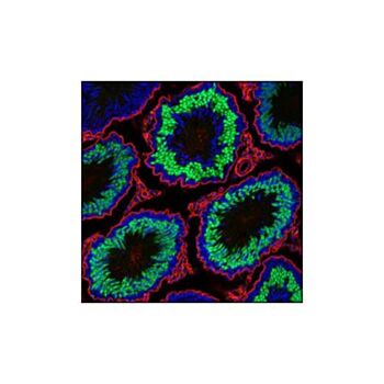
| Application/Dilution | {Western Blotting: 1:1000, Immunoprecipitation: 1:50, Immunohistochemistry (Paraffin): 1:50 - 1:200, Immunofluorescence (Frozen): 1:800 - 1:1600} |
| Storage | Supplied in 10 mM sodium HEPES (pH 7.5), 150 mM NaCl, 100 µg/ml BSA, 50% glycerol and less than 0.02% sodium azide. Store at –20°C. Do not aliquot the antibody. For a carrier free (BSA and azide free) version of this product see product #64162. |
| Specificity/Sensitivity | NUT (C52B1) Rabbit mAb detects endogenous levels of total NUT protein. The antibody also detects endogenous levels of the BRD4-NUT fusion protein found in NUT midline carcinoma (NMC). |
| Species Reactivity | Human, Rat |
| Source/Purification | Monoclonal antibody is produced by immunizing animals with a recombinant protein corresponding to the human NUT protein. |
| Background | Nuclear protein in testis (NUT) is normally confined to the germ cells of the testis and ovary (1,2). NUT midline carcinoma (NMC) is a recently recognized cancer that is defined by the presence of chromosomal rearrangements involving the NUT gene on chromosome 15q14 (3). In most cases the chromosomal translocation occurs between NUT and BRD4 on chromosome 19, resulting in the formation of a BRD4-NUT fusion protein. In the remaining tumors, variant NUT rearrangements are present involving BRD3, a very close homolog of BRD4. BRD4-NUT and BRD3-NUT encode fusion proteins that appear to contribute to carcinogenesis by blocking epithelial cell differentiation. NMCs, which are aggressive and highly lethal carcinomas, are morphologically indistinguishable from other poorly differentiated carcinomas. Given the limited expression of endogenous NUT protein, this antibody can be used to detect NUT fusion proteins in tissues by immunohistochemistry and immunofluorescence (2). |
| SKU | CSIG-3625S |
|---|---|
| Featured | No |
| Supplier Part Number | 3625S |
| UM | EA |
| UNSPSC | 12352203 |
| Manufacturer | Cell Signaling |
| MSDS URL | Click here |
| Temperature | -20C |
| CountryOfOrigin | United States |
| ProductLine | CSIG |
| Qty | 1 |
| MinOrderQty | 1 |
| Weight | 7.000000 |
| Lead Time | 5 |
| Hazardous | N |
| ACT Ecolabel | No |