We use cookies to make your experience better. To comply with the new e-Privacy directive, we need to ask for your consent to set the cookies. Learn about our cookie policy.

Cell Signaling 5-Carboxylcytosine (5-Cac) (D7s8u) Rabbit mAb
List Price
$415.11
Your Price
$415.11
Cell Signaling 5-Carboxylcytosine (5-Cac) (D7s8u) Rabbit mAb - CSIG (Additional S&H or Hazmat Fees May Apply)
NETA PART:
CSIG-36836S
MFG.PART:
36836S
UNSPSC:
12352203
Manufacturer:
Cell Signaling


| Size | 100 µl |
| Reactivity | All |
| Sensitivity | Transfected Only |
| Source/Isotype | Rabbit IgG |
| Application/Dilution | {Immunofluorescence (Immunocytochemistry): 1:200, DNA DOT Blot: 1:1000} |
| Storage | Supplied in 10 mm sodium HEPES (pH 7.5), 150 mm NaCl, 100 µg/mL BSA, 50% glycerol and less than 0.02% sodium azide. Store at –20°C. Do not aliquot the antibody. |
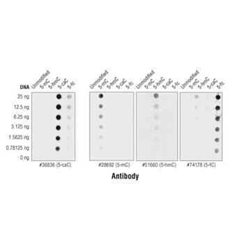
| Specificity/Sensitivity | 5-Carboxylcytosine (5-caC) (D7S8U) Rabbit mAb detects 5-caC by IF in cells over-expressing the TET1 catalytic domain and by DOT blot using double-stranded PCR fragments containing 5-caC. Many cells and tissues contain very low endogenous levels of 5-caC that may fall below the detection limits of this antibody. This antibody has been validated using ELISA, DOT blot, and synthetic spike-in DNA MeDIP assays and shows high specificity for 5-caC. |
| Species Reactivity | All Species Expected |
| Source/Purification | Monoclonal antibody is produced by immunizing animals with 5-carboxylcytidine. |
| Background | Methylation of DNA at cytosine residues is a heritable, epigenetic modification that is critical for proper regulation of gene expression, genomic imprinting, and mammalian development (1,2). 5-methylcytosine is a repressive epigenetic mark established de novo by two enzymes, DNMT3a and DNMT3b, and is maintained by DNMT1 (3, 4). 5-methylcytosine was originally thought to be passively depleted during DNA replication. However, subsequent studies have shown that Ten-Eleven Translocation (TET) proteins TET1, TET2, and TET3 can catalyze the oxidation of methylated cytosine to 5-hydroxymethylcytosine (5-hmC) (5). Additionally, TET proteins can further oxidize 5-hmC to form 5-formylcytosine (5-fC) and 5-carboxylcytosine (5-caC), both of which are excised by thymine-DNA glycosylase (TDG), effectively linking cytosine oxidation to the base excision repair pathway and supporting active cytosine demethylation (6,7).TET protein-mediated cytosine hydroxymethylation was initially demonstrated in mouse brain and embryonic stem cells (5, 8). Since then this modification has been discovered in many tissues, with the highest levels found in the brain (9). While 5-fC and 5-caC appear to be short-lived intermediate species, there is mounting evidence showing that 5-hmC is a distinct epigenetic mark with various unique functions (10,11). The modified base itself is stable in vivo and interacts with various readers including MeCP2 (11,12). The global level of 5-hmC increases during brain development, and 5-hmC is enriched at promoter regions and poised enhancers. Furthermore, there is an inverse correlation between levels of 5-hmC and histone H3K9 and H3K27 trimethylation, suggesting a role for 5-hmC in gene activation (12). Lower amounts of 5-hmC have been reported in various cancers including myeloid leukemia and melanoma (13,14). |
| SKU | CSIG-36836S |
|---|---|
| Supplier Part Number | 36836S |
| UM | EA |
| UNSPSC | 12352203 |
| Manufacturer | Cell Signaling |
| MSDS URL | Click here |
| Temperature | -20C |
| CountryOfOrigin | United States |
| ProductLine | CSIG |
| Qty | 1 |
| MinOrderQty | 1 |
| Weight | 7.000000 |
| Lead Time | 5 |
| Hazardous | N |
| Energy Star | No |
| Green | No |
| Controlled | N |